PCSK9 Inhibitors

comments Comments Off on PCSK9 Inhibitors
By , April 25, 2015 2:31 pm

PCSK9 inhibitors: a major advance in cholesterol-lowering drug therapy

Gregory Curfman, MD, Editor in Chief, Harvard Health Publications

Every so often a medical advance comes along that rewrites the script for treating a disease or condition. After today’s announcements of impressive results of a new type of cholesterol-lowering drug, that scenario just might happen in the next few years.

The new drugs, called PCSK9 inhibitors, are monoclonal antibodies. They target and inactivate a specific protein in the liver. Knocking out this protein, called proprotein convertase subtilisin kexin 9, dramatically reduces the amount of harmful LDL cholesterol circulating in the bloodstream. Lower LDL translates into healthier arteries and fewer heart attacks, strokes, and other problems related to cholesterol-clogged arteries.

http://cen.acs.org/articles/93/i13/Cholesterol-Lowering-PCSK9-Inhibitors-Near.html

CONVERTER mmol to mg/dL conversions for HDL, LDL and cholesterol

comments Comments Off on CONVERTER mmol to mg/dL conversions for HDL, LDL and cholesterol
By , April 22, 2015 4:14 pm

HDL,LDL, cholesterol
mmol/L to mg/dL    /    by 0.0259 to get mg/dL
mg/dL to mmol/L    *    by 0.0259 to get mmol/L

HDL,LDL, cholesterol
mmol/L to mg/dL    /    by 0.0259 to get mg/dL
mg/dL to mmol/L    *    by 0.0259 to get mmol/L

Triglycerides
mmol/L to mg/dL    /    by 0.01129 to get mg/dL
mg/dL to mmol/L    *    by 0.01129 to get mmol/L

EXAMPLE 1 DAD

Cholesterol          4.2  mmol/L    162.16 mg/dL
Triglyceride          0.8 mmol/L    70.85  mg/dL
HDL                  1.11 mmol/L    42.85  mg/dL    needs to be >60mg/dL
LDL                   2.7 mmol/L    104.24 mg/dL     needs to be <70mg/dL
Chol/HDL Ratio       3.8  mmol/L


EXAMPLE 2 MUM

Cholesterol          7.6 mmol/L     293.43 mg/dL
Triglyceride         1.7 mmol/L     150.57 mg/dL
HDL                 1.71 mmol/L      66.02 mg/dL      needs to be >60mg/dL
LDL                  5.1 mmol/L    196.91 mg/ dL     needs to be <70mg/dL
Chol/HDL Ratio       4.4 mmol/L


LDL’s
Bad Fats and promote atherosclerosis (narrowing of the arteries)
An LDL level of less than 100 mg/dL is optimal for CAD prevention,
A level of 70 mg/dL or less is now recommended for persons with existing heart disease
HDL’s
Good Fats and promote opening up of the arteries
> 50 mg/dL (≈ 1.3 mmol/L) man or >60 mg/dL (≈ 1.6 mmol/L) in a woman a reduced risk of atherosclerosis.
>75 mg/dL (≈ 2 mmol/L) man or woman is associated with a very low risk of atherosclerosis.
Less than 40 mg/dL (≈ 0.8 mmol/L) in a man <50 mg/dL (≈ 1 mmol/L) in a woman increases the risk.
TRIGLYCERIDES
Reduction of triglycerides to 60 mg/dl.


HOW TO LOWER YOUR LDL AND CHOLESTEROL

comments Comments Off on HOW TO LOWER YOUR LDL AND CHOLESTEROL
By , April 22, 2015 4:07 pm

LOWER YOUR LDL AND CHOLESTEROL

After reviewing many journals and articles I observe the following ways to lower cholesterol

OBJECTIVES

Important
a) Reduction of LDL to 60 mg/dl (Bad Fats)

An LDL level of less than 100 mg/dL is optimal for CAD prevention,

and a level of 70 mg/dL or less is now recommended for persons with existing heart disease

  1. b) Reduction of triglycerides to 60 mg/dl.
  2. c) Raising HDL to 60 mg/dl. (Good Fats)

> 50 mg/dL (≈ 1.3 mmol/L) man or >60 mg/dL (≈ 1.6 mmol/L) in a woman a reduced risk of atherosclerosis.
>75 mg/dL (≈ 2 mmol/L) man or woman is associated with a very low risk of atherosclerosis.
Less than 40 mg/dL (≈ 0.8 mmol/L) in a man <50 mg/dL (≈ 1 mmol/L) in a woman increases the risk.

  1. d) Achieving normal blood pressure (<130/80)

Even a small elevation of blood pressure in diseased arteries can cause increased mortality.

Diseased arteries are fragile and plaque rupture can occur easily.

  1. f) Achieving normal blood sugar if Diabetic (=100 mg/dl). Diabetes is a high risk factor for heart disease.
  2. g) Reduction of C-reactive protein to <1 mg/l also called (CRP on blood test)


THINGS TO DO

  1. Smoking Cessation
    Is associated with a 200% increase in the rate of atherosclerosis
  2. exercise 45 min cardio daily
  3. Treat underlying conditions:
    Hypertension – Olmesartan medoxomil (angiotensin II receptor antagonist these are preferred)
    Diabetes
    High Cholesterol or LDL ie Take a Statin – medication to lower LDL and cholesterol preferred is Crestor (Rosuvastatin) take at night time (as cholesterol is synthesized by the body at night-time)
  4. Aspirin 100mg / d
  5. Plant Sterols, margarine, 2 g/d The mean one-year reduction in serum cholesterol was 10.2 percent
  6. Dietary Modification:
    1. Mediterranean Diet
    2. Cook with olive oil and use Polyunsaturated fats
    3. Avoid milk fats
    4. Decrease red meat increase fish
    5. Avoidance of fried foods and fast food
  7. Omega 3 (salmon oil) (specifically focusing on high concentration of high in DHA, EPA where possible >2000 mg per day)
  8. Garlic 1g / d
  9. Vit B3 500-1500mg. per day (cheap flush version not anything labelled non-flush which contains inositol)
  10. Wine—Red wines contain resveratrol, (don’t exceed two glasses/ day else has a inverse effect). Bioflavonoids and anti-oxidants have a strong anti-inflammatory effect.
  11. Lechathin
  12. Magnesium
  13. L-carnitine
  14. Vit B6


Fats


Vegetarians and LDL levels

comments Comments Off on Vegetarians and LDL levels
By , April 22, 2015 11:48 am

In epidemiologic studies, a low serum LDL-C of less than 75 mg/dL (≈ 2 mmol/L), typically found in vegetarians and those on non-Western style diets, protects against atherosclerotic cardiovascular disease. Dietary saturated fat and cholesterol generally raise the level of LDL-C in the blood and proportionally raise the coronary risk. The related factors of obesity, metabolic syndrome, and type 2 diabetes may have only modest effects on LDL-C levels, but they increase coronary risk by increasing triglycerides and LDL particle concentration, while lowering HDL-C and causing the production of small, dense LDL particles.
• An HDL level of 50 mg/dL (≈ 1.3 mmol/L) or greater in a man or 60 mg/dL (≈ 1.6 mmol/L) in a woman is associated with a reduced risk of atherosclerosis.
• An HDL level of over 75 mg/dL (≈ 2 mmol/L) or greater in a man or woman is associated with a very low risk of atherosclerosis.
• An HDL of less than 40 mg/dL (≈ 0.8 mmol/L) in a man and less than 50 mg/dL (≈ 1 mmol/L) in a woman increases the risk of atherosclerosis.
• Low HDL levels appear as an independent risk factor for CV events when combined with high LDL levels.


  • LDL Cholesterol – Primary Target of Therapy
    <100 Optimal
    100-129 Near Optimal/Above Optimal
    130-159 Borderline High
    160-189 High
    greater than or equal to   190
    Very high
  • Total Cholesterol
    <200 Desirable
    200-239 Borderline High
    greater than or equal to  240
    High
  • HDL Cholesterol
    <40 Low
    greater than or equal to  60
    High


Cholesterol Risk Categories.png


LDL

The evidence that high LDL concentrations play a causal role in coronary artery disease (CAD)is quite compelling.

  • Many investigators have convincing demonstrated a progressive increase in risk that correlates directly with LDL concentrations.
  • Treatments that lower LDL cholesterol reduce the risk of CAD.
  • Genetic studies analyzing SNPs show that SNPs affecting LDL cholesterol are consistently related to risk of myocardial infarction. In other words, genetic variants associated with elevated LDL levels are consistently associated with increased risk.
  • The presence of LDL in atherosclerotic plaques provides additional evidence of its direct involvement.

Statin Drugs

HMG-CoA-reductase.png 
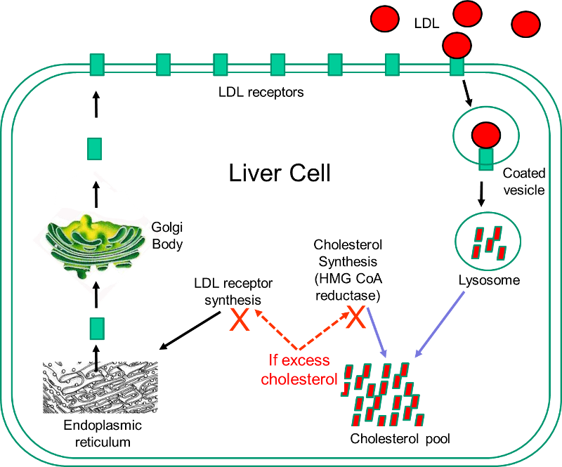
Statins are currently the most powerful cholesterol-lowering drugs available. They act by inhibiting the action of 3-hydroxy-3-methyl-glutaryl-coenzyme A (HMG-CoA) reductase, which is the rate-limiting enzyme in the sequence of steps by which cholesterol is synthesized in the liver. The liver has two sources of cholesterol: it can take up LDL particles from the blood, or it can synthesis cholesterol using HMG-CoA reductase. The diagram to the right illustrates cholesterol homeostasis in a liver cell. LDL (shown in red) can bind to LDL receptors on the surface of the liver. Binding causes the LDL to be taken up by the liver cell and digested in a lysosome. Fatty acids and amino acids in the LDL are recycled, and the cholesterol enters the cholesterol pool in the liver cell. Note that if the liver cell synthesizes cholesterol, this too will be added to the cholesterol pool. If the concentration of cholesterol in the liver cells exceeds a certain level, HMG CoA reductase is inhibited, and the synthesis of LDL receptors is also inhibited.

Statins act by inhibiting HMG-CoA reductase and shutting off internal cholesterol production. In this way, statins reduce cholesterol concentrations in liver cells, which causes increased production of LDL receptors and increased uptake of LDL by the liver. This ultimately results in a lowering of blood concentrations of LDL cholesterol, and this generally results in a slower progression of atherosclerotic plaques and a reduced risk of plaque rupture. However, observers occasionally report regression of atherosclerotic plaques in patient on statins.

While statins are effective in reducing LDL cholesterol levels, they can be associated with side effects.

  • Muscle damage manifest as pain, tiredness, or weakness is the most common side effect and ranges from mild discomfort to difficulty in climbing stairs or walking. Statins very rarely cause severe, life-threatening rhabdomyolysis which can result in severe muscle pain, liver damage, kidney failure, and death.
  • Liver damage: Statins can also damage liver cells, resulting in elevated levels of liver enzymes in blood. Liver enzyme levels should be monitored when statins are first started.
  • Digestive problems: Some people experience nausea, gas, diarrhea or constipation.
  • Rash or flushing
  • Increased blood sugar or type 2 diabetes is occasionally seen in patients on statins.

Because of their effectiveness in reducing CAD in patients at high risk, some advocated expanding the use of statins to people who did not necessarily have elevated cholesterol levels. A number of large, well-done clinical trials have demonstrated that statins can reduce the risk of CAD in low-risk individuals, but most experts caution against this because of the cost and the risk of side effects.


ACE inhibitors decrease AMI risk and are antithrombogenic

comments Comments Off on ACE inhibitors decrease AMI risk and are antithrombogenic
By , April 9, 2015 7:16 pm

As previously described, ACE inhibition may modify not only atherogenesis and plaque vulnerability but also triggering mechanisms responsible for disease onset.229 For example, the renin-angiotensin system may interact with fibrinolytic function,254 and ACE inhibition may influence endogenous fibrinolysis, resulting in a reduced thrombotic response to plaque disruption.255 Importantly, ACE inhibition also seems to reduce mortality and reinfarction in the presence of β-blocker therapy, suggesting an independent therapeutic effect.228

ACE activity may contribute to the development of coronary artery disease and myocardial infarction,225 and ACE inhibition seems to reduce the risk of major ischemic events (reinfarction, cardiac death, and possibly unstable angina) by about 22% in patients with low ejection fractions,226 227 228 probably via multiple beneficial mechanisms.229 ACE inhibitors may influence both atherogenesis (plaque vulnerability) and triggering mechanisms responsible for disease onset (plaque disruption, thrombosis, and/or vasospasm). The latter are discussed below in the section on trigger reduction. The hypothesis that these drugs are antiatherogenic and prevent or slow progression of coronary artery disease is now being tested in clinical trials.

Track Your Plaque

comments Comments Off on Track Your Plaque
By , April 9, 2015 7:03 pm
Track Your Plaque


How to Reverse Heart Disease with the Coronary Calcium Score

by Jeffrey Dach MD


Finally Accepted by the AHA

The AHA (American Heart Association) has steadfastly denied for many years that Coronary Calcium Scoring was a valid marker of heart disease.  Well guess what? They have recanted, and admitted that the amount of calcium in the coronary arteries reliably predicts heart attack risk. This is called the calcium score.(1)

Dr Matt Budoff

UCLA cardiologist, Dr. Matt Budoff, a long-time champion of the Coronary Calcium Scan, and author of the AHA paper says, “The total amount of coronary calcium (Agatston score) predicts coronary disease events beyond standard risk factors.”(1)

Image upper left, courtesy of Wikipedia Rembrandt, The Anatomy Lesson.

Dr. Detrano

Dr. Detrano, in a recent article in NEJM (New England of Medicine), confirms that “The coronary calcium score is a strong predictor of incident coronary heart disease and provides predictive information beyond that provided by standard risk factors”. (31)  The Coronary Calcium Score is a precise quantitative tool for measuring and tracking heart disease risk, and is more valuable and accurate than other traditional markers (such as total cholesterol which is practically worthless as a heart disease risk marker).

What is Coronary Artery Disease?  It’s Plaque Formation.

Coronary Arteries in Cross Section


Age 20-30 years                          Age 50-70 years
In youth, at left, there is minimal plaque formation.  However, at right with passage of time the plaque grows larger. About 20% of this plaque volume contains calcium which is measurable on CAT scan, providing a marker for the total plaque burden.  Calcium score and by inference, plaque volume typically increases 30-35% per year in untreated patients.

Note that even though the right vessel has a larger plaque, the lumen has remodeled so that the inner diameter remains freely open.  Eventually, as we age, the enlarging plaque eventually obstructs blood flow causing a heart attack.  Another common scenario is plaque rupture which exposes the inflammatory debris of the plaque to the circulating blood.  This quickly results in clot formation (thrombosis) resulting in a heart attack and possibly sudden death.

Repeat: The calcified portion of the plaque is consistently 20% of the total plaque volume, allowing use of the calcium score as a marker for total plaque volume.


Arterial Calcification – Why Does it Happen?
Image below: Microscopic view of arterial calcification
(yellow arrows outline blue calcifications)


Calcification in the soft tissues (connective tissue, ligaments, muscles, arteries) is found in many disease states, and commonly identified on pathology slides of tissues.  Whenever there is cell death or tissue necrosis (death of cells), the body invokes a process of calcification which can be regarded as part of the healing process.  Arterial calcification is actually a form of bone formation in the wall of the artery triggered by an inflammatory process.  Pathology studies have shown that coronary artery calcium forms in areas of healed plaque ruptures. (21) Calcification and plaque formation increases with age, with calcium score typically increasing 30-35% per year in untreated patients (William Davis MD).

Heart Attack Rate Associated with Calcium Score


Chart below shows increasing heart attack rate as coronary calcium goes up

Chart Above shows that Calcium Score is Highly Predictive of Heart Attack Risk

What is a Heart Attack?

A heart attack is cell death of heart muscle caused by lack of blood flow with  oxygen deprivation.  AS previously mentioned, this is caused by a arterial blockage by enlarging plaque formation which occludes the lumen, or plaque rupture which causes clot formation which occludes blood flow.  If a small area of heart muscle is involved, the heart attack may be silent with no symptoms.  If a large area is involved, there may be severe chest pain radiating to the left arm or jaw, or other symptoms such as shortness of breath.  If the conduction system is involved, there may be irregular heart rhythm called ventricular tachycardia which can cause sudden death.  Some people have chronic chest pain from diseased arteries and this is called angina pectoris, treated with medicines to dilate the arteries such as nitroglycerine.

Common Sites of Plaque Formation – Bifurcations and Mechanical Stress


(image at right courtesy of Wikipedia)
Above Left image shows xray angiogram of typical ulcerated plaque with stenosis at carotid bifurcation, Above Right Image shows gross pathology of inside of the vessel with darkened plaque (arrow). In this example, we have an artery in the neck that feeds blood flow to the brain.  Plaque rupture and occlusion of the artery in this case caused a stroke, however, the same process occuring in the heart causes a heart attack.

Branching Vessels – Increased Turbulence

Ask any interventional radiologist or invasive cardiologist where they find the plaque formation and obstructions in the arterial tree, and they will say its the same few places over and over again.  These places are the carotid bifurcation, the distal aorta at the bifurcation, the femoral bifurcation, the exit from the adductor canal. And of course, the proximal coronary arteries, and bifurcations of the coronary arteries. A birfurcation is where the vessel branches into two vessels, making a Y pattern.

The bifurcations have maximal turbulance and mechanical stress on the vessel wall.  Remember the blood is flowing under pulsatile pressure, and this mechanical pressure and turbulence, over time, causes little stress cracks in the vessel. The cracks appear at sites of maximal stress.  The coronary arteries are a special case because of the extra motion of the cardiac muscle which moves and stretches the coronary arteries every heart beat, especially as the arteries branch off from the aorta which is relatively stationary, while lower down over the surface of the heart, the vessels move vigorously with each heart beat.  Atherosclerosis is essentially the net result of the healing process for these little cracks in the arterial wall resulting from mechanical stress.

William Davis MDWilliam Davis MD

Advocate of the Coronary Calcium Score

William Davis MD recommends screening CAT heart scans in males over 40 and females over 50.  He would start at younger ages if high-risk features are present, such as strong family history of early heart disease, cigarette smoker, diabetes mellitus, or severe lipid or lipoprotein genetic disorders. (2)(3) The Coronary Calcium Score test is currently covered by Medicare and many health insurances.(32)

Credit and Thanks is given to William Davis MD at the Track Your Plaque Web site for much of the information in this article.  I have added and embellished some of the information.


What does Calcium Look Like?
Normal                  Calcified

Calcium looks White on the CAT scan (red arrows at right).
(Above) CAT scans of the Coronary Arteries.

The left image shows a normal coronary artery (red arrows), while the right image shows a heavily calcified coronary artery (white line outlined by red arrows) indicating high risk for coronary artery disease and heart attack.

All About Coronary Calcium Scoring

 1) Calcium scoring may be superior to angiography as a means to track plaque. That’s because the vast majority of heart attacks are due to plaque rupture and thrombosis at areas of thickened plaque with minimal lumen narrowing.  Over time, the body’s healing process automatically remodels the areas of thickened plaque, and increases lumen size to compensate for the reduced blood flow.

2) Calcium scoring gives a precise number which correlates with the amount of plaque volume. Although only the hard plaque, or calcium in the artery is actually measured, this is useful because it consistently occupies 20% of plaque volume (Total hard and soft plaque).

3) The new 64-slice CAT scanners provide reliable calcium scoring just like any other scanner, both multi-slice and EBT(Electron Beam Cat).


The Track Your Plaque Program, by William Davis MD
(2)(3)

1) Quantify plaque with Coronary Calcium Score with CAT scan (or with Electron Beam CT). Obtain your CAT Scan serially, every 12 months to assess response to treatment and lifestyle modification (track your plaque).

2) Use Sophisticated Lipoprotein Panel (Quest-Cario-IQ , LabCorp-VAP)(7-8)) to uncover hidden causes of plaque progression. LDL particle size and number, Lipoprotein (a). Repeat every 6 months.

3) The Main Treatment Goal is the reduction in Coronary Artery Calcium Score, and by inference, reduction in plaque volume and reduction in cardiovascular mortality. The cardiology community still awaits the hard data on these results (CHD mortality and CHD events, treatment arm vs no treatment arm).  These numbers have not been published as far as I know.

How to Measure Success in Halting or Reversing Heart Disease Plaque

According to Dr. Davis, calcium score typically increases at an astonishing rate of 30-35% per year without treatment. Therefore, Dr. Davis considers treatment success to be reduction in this rate from 30 to perhaps only a 5-10 per cent increase in calcium score per year.  An absolute reduction in calcium score on follow up scanning is the optimal outcome, which is difficult to achieve even with strict adherance to the Track Your Plaque program, in Dr Davis’s experience.

Track Your Plaque Program Details – Attain the Following Targets:

a) Reduction of LDL to 60 mg/dl (LDL should be measured directly, not calculated)

b) Reduction of triglycerides to 60 mg/dl.

c) Raising HDL to 60 mg/dl.

d) Correction of hidden causes of plaque on Lipoprotein profile such as total number of small LDL particles, IDL, and Lp(a).

e) Achieving normal blood pressure (<130/80)  Even a small elevation of blood pressure in diseased arteries can cause increased mortality.  Diseased arteries are fragile and plaque rupture can occur easily.

f) Achieving normal blood sugar (=100 mg/dl). Diabetes is a high risk factor for heart disease.

g) Reduction of C-reactive protein to <1 mg/l

Dietary Modification and Supplements to Attain Above Targets:

Niacin

a) Niacin vitamin B3 (Slo-Niacin Upsher-Smith (44) or Niaspan Kos Pharmaceuticals preferred) 500-1500mg. per day (avoid the no-flush niacin which contains inositol).(6)(44)

Buy Niacin  

on Amazon.

Omega 3 Fish Oil

b) Fish oil (Omega 3 oils) 4000 mg per day (providing 1200 mg omega-3 fatty acids). (molecular distilled pharmaceutical grade).(36)

Buy Purecaps Super-Critical Co2 Extract Omega 3 Fish Oil

Vitamin D

Vitamin D level restored to above 50 ng/ml (Vitamin D3 2000-5,000 u/day), Vitamin K2 also used.  Low vitamin D is associated with increasing arterial calcification and increased heart disease risk. (26)Consumption of calcium tablets by women increases arterial calcification and heart attack risk.(5)

Buy Vitamin D

Read my article on vitamin D which can be found here.(60)

d) Low Glycemic Diet (avoid Fructose Corn Syrup, avoid wheat products), and eliminate wheat products like Shredded Wheat cereal, Raisin Bran, and whole wheat bagels.

e) Consume foods such as raw almonds, walnuts, pecans; olive oil and canola oil. Beneficial for lipoprotein profile.

f) Increasing protein intake, our major building block for body tissues.  Added benefit of protein intake is that it doesn’t increase blood sugar.  This is low glycemic nutrition.

g) Wine—Red wines contain resveratrol, (don’t exceed two glasses/ day). Bioflavonoids and anti-oxidants have a strong anti-inflammatory effect.

h) Fiber – Gound flaxseed (2 tbsp/day)-Extra fiber aids in detoxifying liver and the entire body  by interrupting the enterohepatic circulation. Psyllium (metamucil). Regulates bowel movements and has favorable effect on lipoprotein profile.

Vitamin C – A Genetic Deficiency Disease

Vitamin C (1000–3000 mg/day), is a key player, as it is the vitamin for strong collagen formation, strengthening the arterial wall.  See Linus Pauling’s patented protocol which includes Vitamin C and amino acids Proline and Lysine, the two amino acids that act as receptors for Lp(a).  By consuming additional Lysine and Proline, the receptor sites on the Lp(a) and other lipoproteins are covered up and made less sticky, resulting in less deposition in the artery wall.  The vitamin C is important not only for strong collagen formation, a major component of the arterial wall, but also for all other structural elements of the body, for that matter. (37)(52)(53)(54)(55)(56)(57)

Humans have a genetic deficiency in Gulano-Lactone-Oxidase (GLO), the final enzyme step in the manufacture of Vitamin C, and therefore unlike all the other animals who make their own Vitamin C, we cannot make this necessary vitamin.  We share with all other primates this genetic disease, the inability to manufacture vitamin C, producing a vitamin C deficiency state in all humans.(58)

Also see Thomas Levy’s two books on Vitamin C. (49-51)

Buy Vitamin C at Tower Labs – Ascorcine a9

j) Exercise and weight loss- improves insulin sensitivity, reduces inflammatory markers, reduces blood pressure, improves lipoprotein profile.

Magnesium

k) Magnesium supplementation is inexpensive and safe. Magnesium deficiency due to dietary deficiency or thiazide diuretics for hypertension is common, and is associated increased heart disease risk.  Magnesium reduces blood pressure, relaxes smooth muscle in arteries, and is needed for normal endothelial function.(41-43)

Buy Magnesium

L-Arginine

L-arginine is converted to nitric oxide, an important substance for arterial health. Research by Furchgott and other showed that nitric oxide (NO) relaxes arterial smooth muscle, dilating coronary arteries by up to 50%.(35)  However, Nitric Oxide (NO) is gone after a few seconds, so it must be replenished at a constant rate to keep the arteries relaxed and open. Lack of NO is associated with constricted arteries, damage to the arterial lining, and accelerated plaque growth. L-arginine shrinks coronary plaque,  corrects “endothelial dysfunction”, improves insulin sensitivity, is anti-inflammatory and shrinks plaque.  Dosage: l-arginine 6000 mg twice a day, best taken on an empty stomach.

Buy Arginine

Reverse Cholesterol Transport and Essential Phospholipid – Phosphatidyl Choline (PC) (38)(39)

James C. Roberts MD FACC, a practicing cardiologist, lectures extensively on his clinical success with Phosphatidylcholine (IV or in Liposomal Oral Format with EDTA):  Reverse Cholesterol Transport and Metal Detoxification.  A DVD of his lectures is available which describes considerable clinical success with oral EDTA and phosphytidylcholine.  This page contains his DVD lecture material complete with clinical case histories.(61)

Essential Phospholipid is available under trade name Phoschol from Nutrasol which increases Lecithin Cholesterol Acyl Transferase activity (LCAT) (Dobiasova M 1988).(40)(40b)

Activating LCAT is beneficial because LCAT is the crucial substance which transports cholesterol from the arterial plaque back to the liver for metabolic breakdown into bile.  This process reverses atherosclerotic plaque formation.  Usual Dosage is 3 softgels Nutrasol Phoschol a day each containing 900 mg Phosphatidyl Choline.(38)(39)

Berberine

Berberine, also called Oregon Grape, is a botanical which has many benefits. Dr Roberts page on Berberine for Preventing and Reversing Heart Disease.

Thyroid Function

Normalize thyroid function. Broda Barnes MD showed that low thyroid function was a significant risk factor for heart disease. This conclusion was based on autopsy data from Graz Austria and detailed in his books, Hypothyroidism the Unsuspected Illness, and his other book, Solved the Riddle of Heart Attacks. Barnes felt that the thyroid lab tests were frequently unreliable, and he used clinical judgement instead. (59)

LipoProtein (a)

All About Reducing Lipoprotein (a)(2)(3)

Lipoprotein little A, also written as Lp(a) is a genetic variant lipoprotein which is associated with a high risk of heart disease, and therefore identification and reduction is essential.  The problem is that the conventional Lipid panels done in your doctor’s office do not include Lp(a).  Only the more sophisticated lipoprotein panels such as the Cardio-IQ (Quest), VAP (Atherotech) or NMR (Liposcience) panels provide Lp(a) data.

Lp(a) and Lipoproteins:

1) Lp(a) is best to measured in (nmol/l), and target  below 75 nmol/l .

2) Lp(a) measured in mg/dl (weight may not be accurate), then target below 30 mg/dl .

3) Measured (not calculated) LDL target 50–60 mg/dl.

4) LDL particle number target (NMR) of 600–700 nmol/l or apoprotein B of 50–60 mg/dl. Reduce small LDL to <10% of total LDL.

Treating Lp(a) with Niacin

Use Niaspan® (Kos Pharmaceuticals) or over-the-counter Slo-Niacin® (Upsher-Smith).   Both are better tolerated than OTC plain niacin, which may cause the hot flushes. Reduce hot flushed by drinking a full glass of water with each gelcap, and some find adding an aspirin tablet to the routine helps to reduce flushing.

Lp(a) and BioIdentical Hormones

Bio-Identical hormones are beneficial for reducing heart disease.  In menopausal females, estrogen preparations such as Bi-Est are used. Estrogens have been shown to reduce coronary artery calcium score.(46)

In males over 50, bio-identical testosterone cream may lowers Lp(a) by as much as 25% (per William Davis MD).  Medical studies show that optimizing Testosterone levels in aging males can reduce risk of coronary artery disease by 60%. (47)(48)

DHEA can promote weight loss, and improve insulin sensitivity.(45)


Lp(a) and L-Carnitine

The supplement L-carnitine can be a useful adjunct; 2000–4000 mg per day (1000 mg twice a day) can reduce Lp(a) 7–8%, and occasionally will reduce it up to 20%.

Buy L-Carnitine

Remember, reduction in calcium score on follow up calcium scan is the goal.

What about Statin-Cholesterol Lowering drugs?

Dr Davis admits that the total cholesterol and the LDL cholesterol numbers are of little value in predicting heart disease risk. And he says that the statin drug side effects, ie. muscle pain and weakness, are more common in actual practice than the drug advertising would suggest, making statin drugs difficult to take for the long term.

In my opinion, statin drugs are not recommended for women as explained in my previous article on Statin Drugs for Women, which can be found here (33)

My other article on Statins, Lipitor and the Dracula of Medical Technology can be found here.  (34)

What about Calcium Supplements for women to prevent osteoporosis?

Dr Davis points out that women who take calcium tablets have double the risk of heart attacks than those on placebo.(5)

Check out my earlier Heart Disease Reversal Page here.

Credit and Thanks is given to William Davis MD at the Track Your Plaque Web Site and Blog for the above information.(2)(3)

Fish oil Omega 3 and heart disease

comments Comments Off on Fish oil Omega 3 and heart disease
By , April 7, 2015 1:36 pm

Individuals with high intake of Ω-3 polyunsaturated fatty acids,
usually associated with a high fi sh and low red meat diet, have
low rates of heart disease. Fish oil concentrates reduce triglyceride
levels and increase LDL particle size, a benefi cial effect when
administered in high doses (6–12 g/day).


Patients who had
LDL-C levels of less than 70 mg/dL and CRP levels of less than
1 mg/L after statin therapy had the lowest rate of recurrent CV
events.


Effect of Plant Sterol–Containing Fats on Blood Lipid Levels

comments Comments Off on Effect of Plant Sterol–Containing Fats on Blood Lipid Levels
By , April 7, 2015 1:35 pm

Effect of Plant Sterol–Containing Fats on Blood Lipid Levels

Background

In the early 1950s, plant-derived sterols were observed to decrease serum cholesterol levels.1 2 The effective dose in humans was reported to be between 5 and 10 g/d when given in divided doses. The efficacy of plant sterols with regard to lowering blood cholesterol levels was soon confirmed,3 4 5 6 albeit at somewhat lower doses.7 On the basis of these data, plant sterols were briefly used in the reduction of blood cholesterol levels before the introduction of pharmacological agents with higher efficacy and patient acceptance. The resurgence of interest in plant-derived sterols is now coupled with the incorporation of these compounds into fat-containing foods. More recent evidence has shown that esterification of these sterols increases their solubility in fat and their efficacy in lowering low-density lipoprotein (LDL) cholesterol levels.8 9

What Are Plant-Derived Sterols?

Sterols represent a group of compounds that are alcoholic derivatives of cyclopentanoperhydrophenanthrene and are an essential constituent of cell membranes in animals and plants. Cholesterol is the sterol of mammalian cells, whereas multiple sterols, or phytosterols, are produced by plants, with sitosterol, campesterol, and stigmasterol being most common. Plant sterols, although structurally similar to cholesterol, are not synthesized by the human body. They are very poorly absorbed by the human intestine. The specific plant sterols that are currently incorporated into foods intended to lower blood cholesterol levels are extracted from soybean oil or tall (pine tree) oil. Additional sources of plant sterols may be available in the near future. The plant sterols currently incorporated into foods are esterified to unsaturated fatty acids (creating sterol esters) to increase lipid solubility, thus allowing maximal incorporation into a limited amount of fat. Some plant sterols currently available are saturated, to form the stanol derivatives, sitostanol and campestanol, which after esterification form stanol esters.

Effect of Plant Sterol–Containing Fats on Blood Lipid Levels

A clinical study of hypercholesterolemic subjects concluded that esterified β-sitostanol was more efficient than free β-sitosterol, free β-sitostanol, or rapeseed-based margarine alone in lowering serum cholesterol (14). The superiority of β-sitostanol ester has yet to be confirmed because the intakes of the dietary plant sterols were different in the experimental groups of the trial. A comparison of free β-sitostanol with free β-sitosterol, however, showed that the saturated plant sterol increased cholesterol excretion more effectively than the unsaturated plant sterol when infused over several hours in low concentrations (3). Despite the latter finding, a recent comparison of esterified unsaturated sterols from soybeans with the ester of the saturated β-sitostanol indicated that soybean sterol esters had a similar serum cholesterol–lowering effect as the β-sitostanol ester (12). It could therefore be hypothesized that soy sterols and β-sitostanol inhibit cholesterol absorption equally when both fractions are esterified.

In the early 1990s, it was reported that sitostanol ester (3.4 g/d) delivered in the form of rapeseed (canola) oil–based margarine lowered LDL cholesterol levels by ≈10% in modestly hypercholesterolemic subjects and that individuals with apolipoprotein (apo) E4 alleles, previously reported to have the highest efficiency of cholesterol absorption, derived the greatest benefit from treatment.8 9 Subsequent work has established that maximal efficacy with respect to total and LDL cholesterol lowering is achieved at ≈2 g/d and that there is little or no effect on high-density lipoprotein (HDL) cholesterol or triglyceride levels.10 11 12 13 14 15 16 17 18 19 20 21 In addition, these studies demonstrated that the consumption of fats containing plant-derived sterol esters is efficacious in both normolipidemic and dyslipidemic individuals, including those treated with β-hydroxy-β-methylglutaryl–coenzyme A (HMG-CoA) reductase inhibitors and other lipid-lowering agents. In addition, daily ingestion of 1.8 to 3 g of plant stanol esters in hypercholesterolemic children has been reported to reduce LDL cholesterol levels to an extent similar to that in adults.22 For the most part, the consumption of ≈2 g/d of plant sterol ester has been reported to decrease LDL cholesterol levels 9% to 20%, with considerable variability reported among individuals.10 11 12 13 14 15 16 17 18 19 20 21 There appears to be little difference in efficacy of campestanol ester and sitostanol ester with respect to cholesterol lowering.23

The impact of the sterol/stanol ester–containing fats on LDL cholesterol lowering is relatively consistent regardless of whether the background diet is similar to that currently consumed in Western countries or reduced in total fat, saturated fat, and cholesterol, consistent with current guidelines for hypercholesterolemic individuals, and whether the plant sterol ester–containing fats have been incorporated into regular fat or reduced fat products.8 9 10 11 12 13 14 15 16 17 18 19 20 21

Both unsaturated (sterol ester) and saturated (stanol ester) forms of plant sterols have been used in the above studies. Comparative investigations of the relative efficacy of these 2 preparations in regular-fat margarine have recently been reported.15 21 Superimposed on a background diet high in total and saturated fat, ≈2 to 3 g/d taken in 2 divided doses of both sitostanol ester and sitosterol ester in margarine resulted in 10% to 13% reductions in LDL cholesterol and no significant change in HDL cholesterol levels. An additional study has compared the effect of 2 reduced-fat (40% of fat) margarines containing stanol esters, the sterol esters derived from either tall oil or soybean oil, within the context of diets consistent with the American Heart Association Step 2 diet criteria.24 The efficacy of both preparations was similar, with a decrease of ≈9% in LDL cholesterol levels.

Mechanisms of Action of Plant Stanol/Sterol Ester–Containing Fats

Sterol balance studies have suggested that decreased blood cholesterol levels are attributable, at least in part, to an inhibition of cholesterol absorption.25 This inhibition has been ascribed to a number of mechanisms, including partitioning in the micellar phase of the intestinal lumen, presence in the unstirred water layer or other mucosa barriers that might limit transmembrane transport, and alteration in rates of cholesterol esterification in the intestinal wall.25 26 27 28 29

Plant sterols differ structurally from cholesterol by a methyl or ethyl group in their side chains and are not synthesized in the human body. These structural differences render them minimally absorbable. Serum campesterol levels and stable isotope–labeled cholesterol can be used to estimate the efficiency of intestinal cholesterol absorption in humans.27 28 29 Such data have confirmed the original observations from sterol balance studies that plant-derived sterols decrease the absorption of both dietary and endogenously derived cholesterol in the intestine. It has been speculated that the full magnitude in the decreased rate of cholesterol absorption (33% to 60%) is not realized in decreased LDL cholesterol levels because of compensatory mechanisms that increase the rate of endogenous cholesterol synthesis.8 9 This speculation has recently been confirmed.30 Lipoprotein kinetic studies have associated the significant decreases in LDL cholesterol levels with a decreased production rate of LDL apoB rather than a change in the LDL apoB fractional catabolic rate.12 The general lack of effect of plant-derived sterols on HDL cholesterol levels was reflected in essentially no change in the kinetic parameters of HDL apoA-I.12

Potential Risks Associated With the Use of Plant Stanol/Sterol Ester–Containing Fats

Few adverse effects related to either the short-term or long-term consumption of the plant stanol/sterol ester–containing fats have been reported. However, of concern are some observations of decreased levels of plasma alpha plus beta carotene, α-tocopherol, and/or lycopene as a result of the consumption of foods containing both stanol esters and sterol esters.16 17 23 24 In general, with the exception of beta carotene, these decreases often parallel the decreases in total and LDL cholesterol. Still, at this time it appears prudent to recommend additional monitoring of the effect of foods containing plant-derived sterol/stanol esters on fat-soluble nutrient levels and to recommend that an assessment of the biological significance of the changes observed be determined. The activities of alkaline phosphatase, alanine transaminase, aspartate transaminase, and γ-glutamate transaminase have been reported to be unaffected by plant sterol consumption within the recommended range.17 Other technical data on safety evaluation are now available.31 32 33 34 35 36 37

Plasma levels of plant sterols/stanols have not been or are only minimally elevated after daily ingestion of sterol/stanol ester–containing foods.12 16 23 24 However, there may be some individuals in the population who have abnormally high absorption of plant sterols. For example, individuals homozygous for sitosterolemia absorb substantial amounts of sitosterol, with resultant hypercholesterolemia and development of xanthomas.38 It is not known whether some individuals heterozygous for this condition could absorb higher amounts of plant sterols than the normal population and whether this would lead to adverse effects. In a study of 2 obligate heterozygotes for sitosterolemia,39 increased sitosterol absorption was balanced by enhanced plant sterol elimination. It is not known what percentage of individuals in a given population would have this condition. Still, in the absence of more data on genetic mutations involved in sitosterolemia, it would be prudent to counsel these individuals against the use of these foods at the present time.

Of concern are the potential adverse effects of lowering beta carotene and perhaps other fat-soluble vitamins over long periods of time in children who would be ingesting plant sterol–containing fats. Likewise, data on the effect of these compounds in pregnant women are lacking. Because food products containing plant sterols are likely to be shared during meals by all family members, the potential for intake by nonhypercholesterolemic individuals is significant. Thus, the American Heart Association recommends that further studies and large-scale monitoring be undertaken to determine the long-term safety of plant sterol/stanol ester–containing foods in both normocholesterolemic and hypercholesterolemic adults, as well as in children.

Outcome

The margarine containing sitostanol ester was well tolerated. The mean one-year reduction in serum cholesterol was 10.2 percent in the sitostanol group, as compared with an increase of 0.1 percent in the control group. The difference in the change in serum cholesterol concentration between the two groups was -24 mg per deciliter (95 percent confidence interval, -17 to -32; P<0.001). The respective reductions in low-density lipoprotein (LDL) cholesterol were 14.1 percent in the sitostanol group and 1.1 percent in the control group. The difference in the change in LDL cholesterol concentration between the two groups was -21 mg per deciliter (95 percent confidence interval, -14 to -29; P<0.001). Neither serum triglyceride nor high-density lipoprotein cholesterol concentrations were affected by sitostanol. Serum campesterol, a dietary plant sterol whose levels reflect cholesterol absorption, was decreased by 36 percent in the sitostanol group, and the reduction was directly correlated with the reduction in total cholesterol (r = 0.57, P<0.001).

Modifying Coronary Artery Atherosclerosis

comments Comments Off on Modifying Coronary Artery Atherosclerosis
By , April 7, 2015 11:18 am


Framingham Heart Study Identifying major CVD risk factors

Over the years, careful monitoring of the Framingham Study population has led to the identification of the major CVD risk factors –
high blood pressure,
high blood cholesterol,
smoking,
obesity,
diabetes, and
physical inactivity

Hypertension
Each 20-mm Hg increase in systolic blood pressure or 10-mm Hg increase in diastolic blood pressure doubles coronary heart disease mortality and stroke mortality. Proposed mechanisms by which hypertension promotes atherosclerosis include damage to endothelial cells, promoting activation and incorporation of lipids into subintima, and stimulation of subintimal smooth muscle cell proliferation.


For prevention of CAD, the NCEP has determined that an LDL-C level of less than 100 mg/dL is optimal for CAD prevention, and a level of 70 mg/dL or less is now recommended for persons with existing CAD plus other risk factors. Ideal blood pressure was set at 120/80 mm Hg or less by


low-dose aspirin, and fish oil—may be considered, depending on the age and estimated CV risk of the patient.

Each 20–mm Hg increase in systolic blood pressure or 10–mm Hg increase in diastolic blood pressure doubles coronary artery disease mortality and stroke mortality.

It is instructive to note that although human LDL-C concentrations tend to cluster from 120 to 200 mg/dL and atherosclerotic disease is the main cause of death in humans, typical nonhuman mammalian LDL-C levels are 10 to 60 mg/dL, and atherosclerosis does not occur unless animals are fed high-fat food.


The risk of a coronary event is thought to increase by 2%for every 1%decrease in plasma HDL


Diet and exercise

Certain changes in diet and exercise may have a positive impact on raising HDL levels:[29]

Most saturated fats increase HDL cholesterol to varying degrees and also raise total and LDL cholesterol.[42] A high-fat, adequate-protein, low-carbohydrate ketogenic diet may have similar response to taking niacin (vitamin B3) as described below (lowered LDL and increased HDL) through beta-hydroxybutyrate coupling the Niacin receptor 1.[43]

HDL levels can be increased by smoking cessation,[35] or mild to moderate alcohol intake.[44][45][46][47][48][49]

Pharmacologic (1- to 3-gram/day) niacin doses increase HDL levels by 10–30%,[53] making it the most powerful agent to increase HDL-cholesterol.[54][55] A randomized clinical trial demonstrated that treatment with niacin can significantly reduce atherosclerosis progression and cardiovascular events.[56] However, niacin products sold as “no-flush”, i.e. not having side-effects such as “niacin flush“, do not contain free nicotinic acid and are therefore ineffective at raising HDL, while products sold as “sustained-release” may contain free nicotinic acid, but “some brands are hepatotoxic”; therefore the recommended form of niacin for raising HDL is the cheapest, immediate-release preparation.[57]

AIM-HIGH trial (Atherothrombosis Intervention in Metabolic Syndrome with Low HDL/High Triglycerides

As above, but instead of a placebo, patients were given 1,500 to 2,000 mg/day of extended-release niacin.

After two years the niacin group, as expected, had experienced a significant increase in plasma HDL-C (along with some other benefits like a greater reduction in plasma triglycerides).  However, there was no improvement in patient survival.  The trial was futile and the data and safety board halted the trial.  In other words, for patients with cardiac risk and LDL-C levels at goal with medication niacin, despite raising HDL-C and lowering TG, did nothing to improve survival. This was another strike against the HDL hypothesis.


CETP. – Dalcetrapib

By 2008, as the AIM-HIGH trial was well under way, another pharma giant, Roche, was well into clinical trials with another drug that blocked CETP.  This drug, a cousin of torcetrapib called dalcetrapib, albeit a weaker CETP-inhibitor, appeared to do all the “right” stuff (i.e., it increased HDL-C) without the “wrong” stuff (i.e., it did not appear to adversely affect blood pressure). It did nothing to LDL-C or apoB.  This study, called dal-OUTCOMES, was similar to the other trials in that patients were randomized to either standard of care plus placebo or standard of care plus escalating doses of dalcetrapib.   A report of smaller safety studies (called dal-Vessel and Dal-Plaque) was published a few months ago in the American Heart Journal, and shortly after Roche halted the phase 3 clinical trial.  Once again, patients on the treatment arm did experience a significant increase in HDL-C, but failed to appreciate any clinical benefit.  Another futile trial.


Mendelian randomization

On May 17 of this year a large group in Europe (hence the spelling of randomization) published a paper in The Lancet, titled, “Plasma HDL cholesterol and risk of myocardial infarction: a mendelian randomisation study.”  Mendelian randomization, as its name sort of suggests, is a method of using known genetic differences in large populations to try to “sort out” large pools of epidemiologic data.

In the case of this study, pooled data from tens of studies where patients were known to have myocardial infarction (heart attacks) were mapped against known genetic alterations called SNPs (single nucleotide polymorphisms, pronounced “snips”).  I’m not going to go into detail about the methodology because it would take 3 more blog posts., But, the reason for doing this analysis was to ferret out if having a high HDL-C was (only) correlated with better cardiovascular outcome, which has been the classic teaching, or if there was any causal relationship.  In other words, does having a high HDL-C cause you to have a lower risk of heart disease or is it a marker for something else?

This study found, consistent with the trials I’ve discussed above, that any genetic polymorphism that seems to raise HDL-C does not seem to protect from heart disease.  That is, patients with higher HDL-C due to a known genetic alteration did not seem to have protection from heart disease as a result of that gene. This suggests that people with high or low HDL-C who get coronary artery disease may well have something else at play.


https://s-media-cache-ak0.pinimg.com/originals/13/32/8b/13328b0ba7b1dde753d4564838415f17.jpg

Dr Thomas Challenger Challenger Mission